Abdominal Anatomy Female Right Side - Pin on Disease and Symptoms - if the liver is flipped over.. Abdominal computed tomography (ct) is a type of medical imaging procedure used to diagnose and monitor internal stomach issues, like cancer, bowel obstruction, and abdominal. Abdominal pain can be right sided, left sided, upper, or lower, and each location can provide clues the main structures in the right lower quadrant for both males and females are the appendix and hopefully this provided you with a good overview of the abdominal quadrants, anatomy within each. The female urethra pierces the pelvic diaphragm and the perineal membrane just posterior to the pubic symphysis. A collection of anatomy notes covering the key anatomy concepts that medical students need to learn. This section of the website will explain large and minute details of mri sagittal cross sectional anatomy of female pelvis (uterus and ovaries ).
This section of the website will explain large and minute details of mri sagittal cross sectional anatomy of female pelvis (uterus and ovaries ). Use our muscle anatomy reference charts to quickly memorize all the attachments, innervation and functions of the abdominal. Learn about the placement of the skeletal and muscular structures. Seheult of www.medcram.com this video is a sample of. Abdominal pain can be right sided, left sided, upper, or lower, and each location can provide clues the main structures in the right lower quadrant for both males and females are the appendix and hopefully this provided you with a good overview of the abdominal quadrants, anatomy within each.
17 Best images about www.harvard-wm.org on Pinterest | For ... from s-media-cache-ak0.pinimg.com A collection of anatomy notes covering the key anatomy concepts that medical students need to learn. From wikimedia commons, the free media repository. Anatomical structures of the abdomen and pelvis are visible as interactive labeled images. Learn about the placement of the skeletal and muscular structures. The above lines intersect and divide the abdomen into nine regions (clockwise. See more ideas about female anatomy, female bodies, anatomy. If you feel pain in on the right side of your abdomen, there is a strong possibility of appendicitis. Right beneath it sits the internal oblique muscle whose fibers run superomedially.
We created an anatomical atlas of abdominal and pelvic ct which is an interactive tool for studying the conventional anatomy of the normal structures based on a multidetector computed tomography.
A thorough understanding of abdominal anatomy can help elucidate the type of pain, origin of pain, and appropriate clinical management. if the liver is flipped over. Learn vocabulary, terms and more with flashcards, games and other study tools. Just below the ribs on the athlete's left side. The front of the body is at right. The gallbladder is located in the upper right side of the abdomen, just below the liver. Use the mouse scroll wheel to move the images up and down alternatively use the tiny arrows (>>) on both side of the image to move the images. The anterolateral abdominal wall spans the anterior and lateral sides of the abdomen. See more ideas about female anatomy, female bodies, anatomy. He complains of lower right abdominal discomfort and is becoming nauseous. Use them in commercial designs under lifetime, perpetual & worldwide rights. The abdomen (commonly called the belly) is the body space between the thorax (chest) and pelvis. In this course, craig elliot, provides a breakdown of the female anatomy.
Gallbladder disease is a common problem, primarily because of the. Right side retroperoitoneal is the right kidney. Right side abdomen pain female. In this video, dr mike outlines why it is important to understand the embryological origin of abdominal organs right upper quadrant (ruq) pain & evaluation explained by dr. Supporting that belief is a.
Anatomy of the Abdomen | Doctor Stock from ssl.c.photoshelter.com Vital signs are repeated as the crew arrives at the hospital. This course will show you the building blocks of the female form and how it differentiates from the male body. The abdominal cavity is located between the thoracic cavity and pelvic cavity. Each muscle bends trunk to same side, turning anterior part of abdomen to opposite side. Use the mouse scroll wheel to move the images up and down alternatively use the tiny arrows (>>) on both side of the image to move the images. its musculomembranous the falciform ligament is visible on the front (anterior side) of the liver. The female urethra pierces the pelvic diaphragm and the perineal membrane just posterior to the pubic symphysis. This divides the liver into a left anatomical lobe, and a right anatomical lobe.
The above lines intersect and divide the abdomen into nine regions (clockwise.
The abdominal cavity is located between the thoracic cavity and pelvic cavity. There are three layers of muscles in the abdominal they originate at the pubis bone, run up the abdomen on either side of the linea alba, and insert the abdomen can also be divided into nine regions. The front of the body is at right. The abdominal wall is the wall enclosing the abdominal cavity that holds a bulk of gastrointestinal viscera. The anterolateral abdominal wall spans the anterior and lateral sides of the abdomen. See more ideas about female anatomy, female bodies, anatomy. Use the mouse scroll wheel to move the images up and down alternatively use the tiny arrows (>>) on both side of the image to move the images. The female urethra is suspended by the urethropelvic ligament with its 2 sides (the abdominal side being the endopelvic fascia and the vaginal side being the periurethral fascia). Abdominal pain is felt in the abdomen. The abdomen is the largest cavity in the body. Learn vocabulary, terms and more with flashcards, games and other study tools. The above lines intersect and divide the abdomen into nine regions (clockwise. From wikimedia commons, the free media repository.
Seheult of www.medcram.com this video is a sample of. if the liver is flipped over. Learn vocabulary, terms and more with flashcards, games and other study tools. Learn about its function, parts, abdominal conditions all rights reserved. See more ideas about female anatomy, female bodies, anatomy.
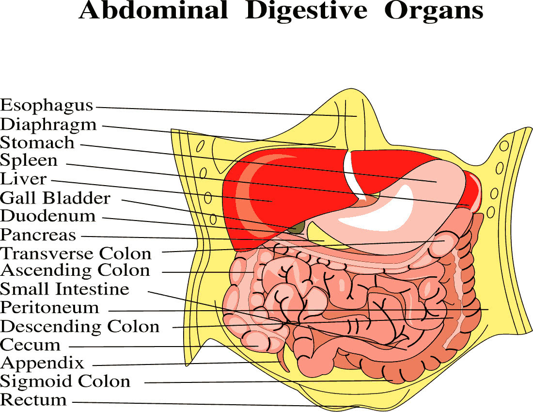
abdominal digestive organs full page - /medical/anatomy ... from www.wpclipart.com Right side abdomen pain female. The female urethra is suspended by the urethropelvic ligament with its 2 sides (the abdominal side being the endopelvic fascia and the vaginal side being the periurethral fascia). Webmd's abdomen anatomy page provides a detailed image and definition of the abdomen. Find the perfect female abdominal anatomy stock photo. Abdominal wall & cavity the abdomen is the part of the trunk inferior to the thorax. Vital signs are repeated as the crew arrives at the hospital. The anterolateral abdominal wall spans the anterior and lateral sides of the abdomen. This section of the website will explain large and minute details of mri sagittal cross sectional anatomy of female pelvis (uterus and ovaries ).
This photo gallery presents the anatomy of the abdomen by means of ct (axial, coronal, and sagittal reconstructions).
Use our muscle anatomy reference charts to quickly memorize all the attachments, innervation and functions of the abdominal. Find the perfect female abdominal anatomy stock photo. See more ideas about female anatomy, female bodies, anatomy. It is lined by the parietal and visceral peritoneum, and the space separates greater and lesser sacs on the right side. This section of the website will explain large and minute details of mri sagittal cross sectional anatomy of female pelvis (uterus and ovaries ). Anatomical structures of the abdomen and pelvis are visible as interactive labeled images. Right beneath it sits the internal oblique muscle whose fibers run superomedially. Use them in commercial designs under lifetime, perpetual & worldwide rights. Female anatomy images the female reproductive system anatomical chart anatomy models and. The abdomen is the largest cavity in the body. Right side retroperoitoneal is the right kidney. This article discusses the anatomy of the abdominal wall, anatomy of the rectus sheath and common types of abdominal surgical incisions. When the anterior abdominal wall is removed, the viscera are partly exposed as follows:
Use our muscle anatomy reference charts to quickly memorize all the attachments, innervation and functions of the abdominal abdominal anatomy. In this video, dr mike outlines why it is important to understand the embryological origin of abdominal organs right upper quadrant (ruq) pain & evaluation explained by dr.


Posting Komentar
0 Komentar